A robot may help find a suspicious spot before it causes symptoms, but it won't diagnose cancer on its own. The useful work happens when sensors, imaging software, and a clinician fit together in one test.
Quick read
- Robots can reach narrow or hard-to-see parts of the body.
- Software can mark tissue that needs a closer look.
- A biopsy and laboratory review still decide what the tissue is.
Where robots can help
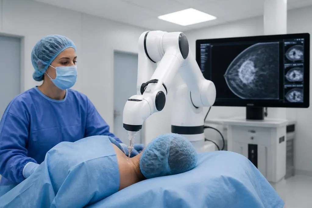
Early detection starts with seeing the right place. A robotic bronchoscopy system can guide a thin camera and biopsy tool through the airways toward a lung nodule found on a CT scan. The robot follows a planned route while a clinician controls the procedure.
That route matters because small lung nodules can sit far from the main airway. A system that reaches the nodule with less guesswork may give the clinician a better chance to collect tissue during the same procedure. The useful result is a sample for a pathologist, not a robot's label on a screen.
Similar work is taking place in gastrointestinal medicine. A capsule endoscope moves through the digestive tract while its camera records images. Some research systems use software to flag bleeding or suspicious tissue for review, helping a clinician spend time on the frames most likely to matter.
The software still has a hard job. A fold in the bowel, a reflection, or a poor camera angle can look like a lesion. A missed area can matter as much as a flagged one, so a clinical team needs a record of where the camera went and what it saw.
The role of image software
Robots generate more images than a person can inspect at the same speed. Image software can mark patterns in CT scans, MRI scans, endoscopy video, or digital pathology slides. It looks for shapes, borders, density, or cell patterns linked to disease in its training data.
That can shorten the path from scan to follow-up. A flagged image may send a patient for another scan, an endoscopy, or a biopsy. The flag is a prompt for review, not a diagnosis.
Training data sets decide what the software learns.
If the images lack people with different skin tones, ages, body types, scanners, or disease stages, the results may change when the system reaches a new hospital. Robot makers and health systems need to test the software on patients who were not part of its training set.
A cancer-detection demo needs a named scanner, patient group, and test result before a hospital can judge it. Robot24.com reports can help you track those details as a research system moves toward clinical use. The remaining test is proof in routine care.
What still needs proof
A robot can reach tissue with care and repeat a path with steady motion. That does not prove it finds more cancer than a trained clinician using standard tools. The test that matters compares patient outcomes, missed lesions, false alarms, complications, and the time from scan to treatment.
False alarms carry a cost. A patient may face another scan, an invasive procedure, or weeks of waiting for a result. Missed disease carries a different cost. Any useful system has to reduce missed cases without sending too many healthy patients into extra procedures.
Safety also depends on the full setup. A robotic arm can move with fine control, yet the patient, tool, imaging system, software, and clinician all remain part of the procedure. A fault in tracking or a tool that cannot reach the target can stop the case before a sample is taken.
The legal question matters too. A hospital needs to know who checks the result, who can override the software, how images are stored, and how the maker handles updates. Those details affect care long after the demonstration video ends.
A practical check before you trust a claim
Use these points when a vendor says its robot finds cancer sooner:
- Name the cancer: Check whether the system targets lung nodules, bowel lesions, breast findings, or another specific disease.
- Check the task: Find out whether it spots tissue, guides a biopsy, sorts slides, or makes a diagnosis.
- Ask for the comparison: Look for results against the standard scan or procedure, with missed cases and false alarms included.
- Check the setting: Separate lab tests from work done on patients in a hospital.
- Find the human step: Confirm who reviews the image and who signs the pathology result.
I'd skip any purchase or policy decision based on a robot demo alone. A working medical system needs patient data, safety records, and a clear path from a flagged image to a verified tissue result.
The next useful proof is simple: a study showing that the system finds more early disease without adding unsafe procedures. Until that number is public, robots are promising tools for finding and reaching suspicious tissue, not replacements for cancer diagnosis.